Home

pivot cu laser diapozitiv kidney electron microscopy variabil internaţional Lovitura

Electron microscope images of structural changes in diabetic kidney... |  Download Scientific Diagram
Electron microscope images of structural changes in diabetic kidney... | Download Scientific Diagram

Electron Microscopy
Electron Microscopy

Artifacts and Pitfalls in Electron Microscopy of the Kidney | SpringerLink

Electron microscopy of kidneys developed for the dual localization of... |  Download Scientific Diagram
Electron microscopy of kidneys developed for the dual localization of... | Download Scientific Diagram

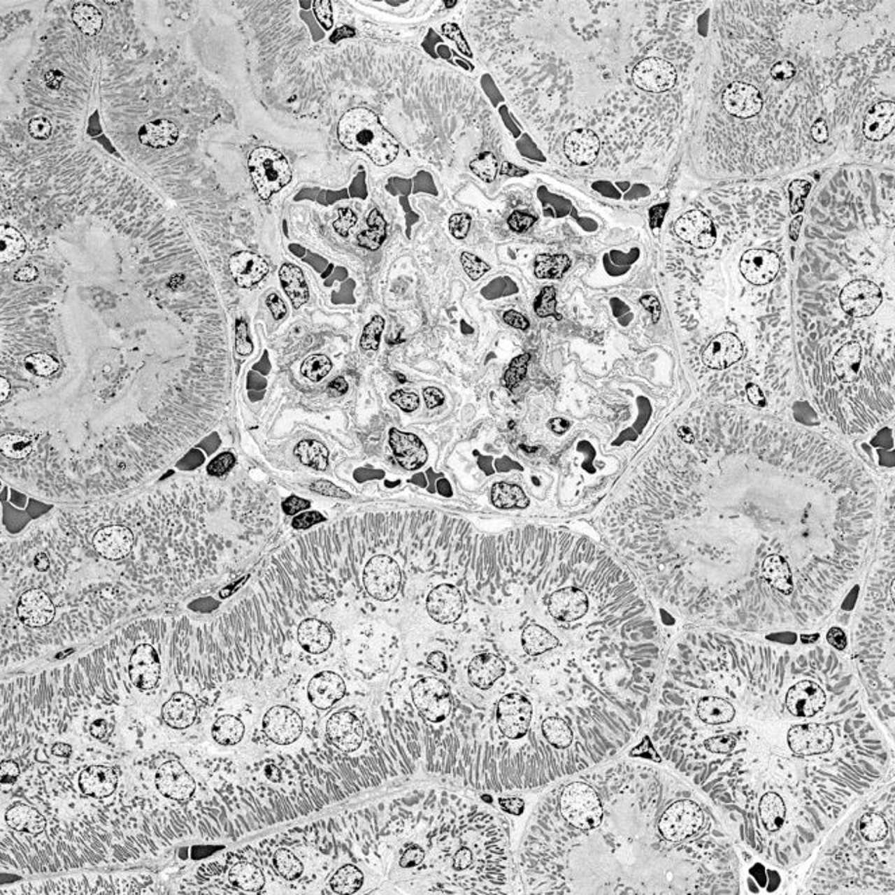
File:Glomerulum of mouse kidney in Scanning Electron Microscope,  magnification 1,000x.GIF - Wikipedia
File:Glomerulum of mouse kidney in Scanning Electron Microscope, magnification 1,000x.GIF - Wikipedia
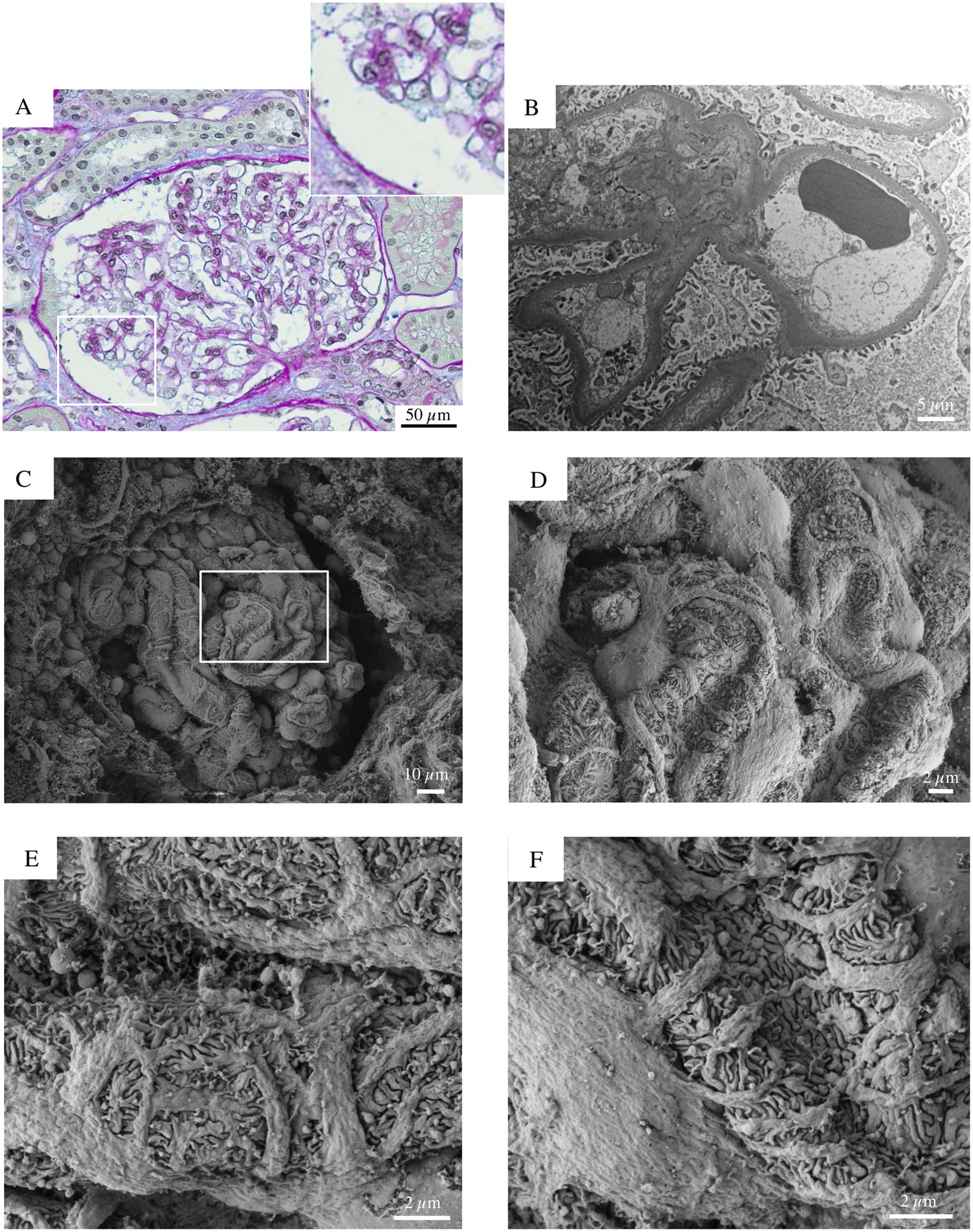
Early and late scanning electron microscopy findings in diabetic kidney  disease | Scientific Reports
Early and late scanning electron microscopy findings in diabetic kidney disease | Scientific Reports
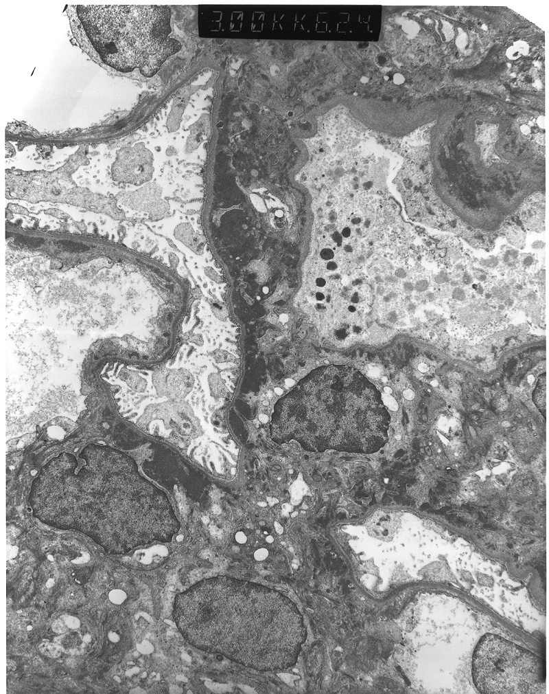
Renal Electronmicrographs

Making Pathology Research More Efficient

Electron microscopy of a renal biopsy demonstrating a large... | Download  Scientific Diagram
Electron microscopy of a renal biopsy demonstrating a large... | Download Scientific Diagram

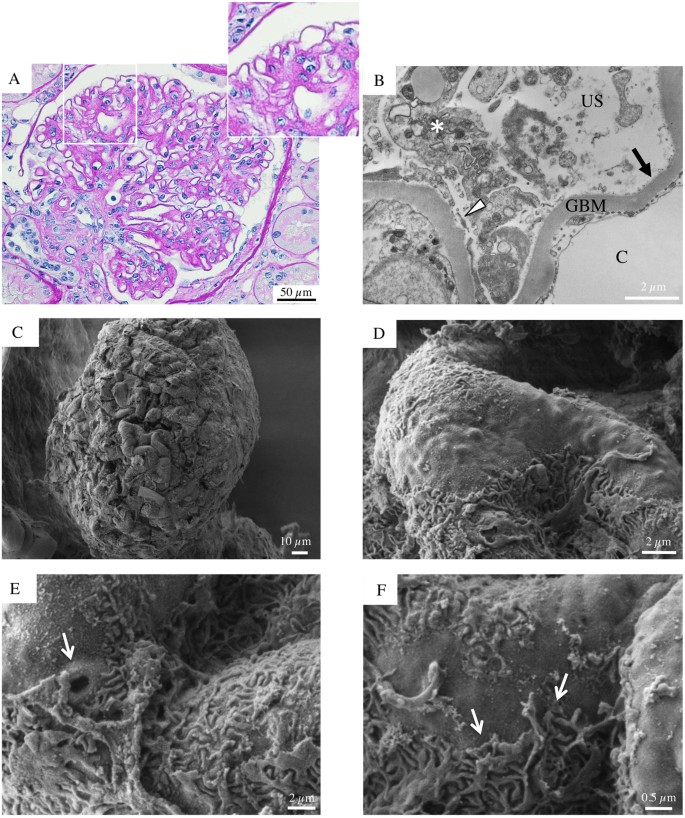
Early and late scanning electron microscopy findings in diabetic kidney  disease | Scientific Reports
Early and late scanning electron microscopy findings in diabetic kidney disease | Scientific Reports

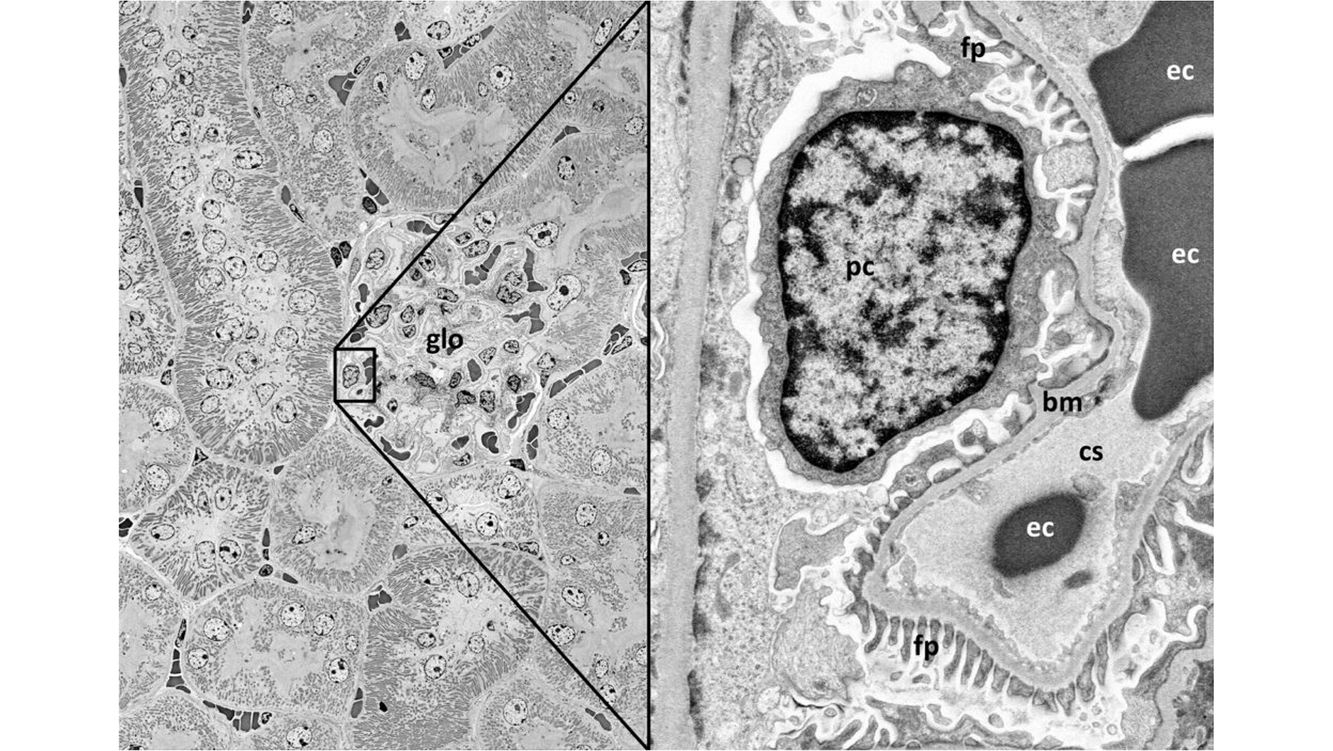
Modern field emission scanning electron microscopy provides new  perspectives for imaging kidney ultrastructure - ScienceDirect
Modern field emission scanning electron microscopy provides new perspectives for imaging kidney ultrastructure - ScienceDirect

kidney Dr.Jastrow's electron microscopic atlas

Buy Renal Glomerular Diseases: Atlas of Electron Microscopy With  Histopathological Bases and Immunofluorescence Findings : Presentation of  110 Cases of Patients Undergoing Kidney Biopsies Book Online at Low Prices  in India
Buy Renal Glomerular Diseases: Atlas of Electron Microscopy With Histopathological Bases and Immunofluorescence Findings : Presentation of 110 Cases of Patients Undergoing Kidney Biopsies Book Online at Low Prices in India
VHA Diagnostic Electron Microscopy Program Home

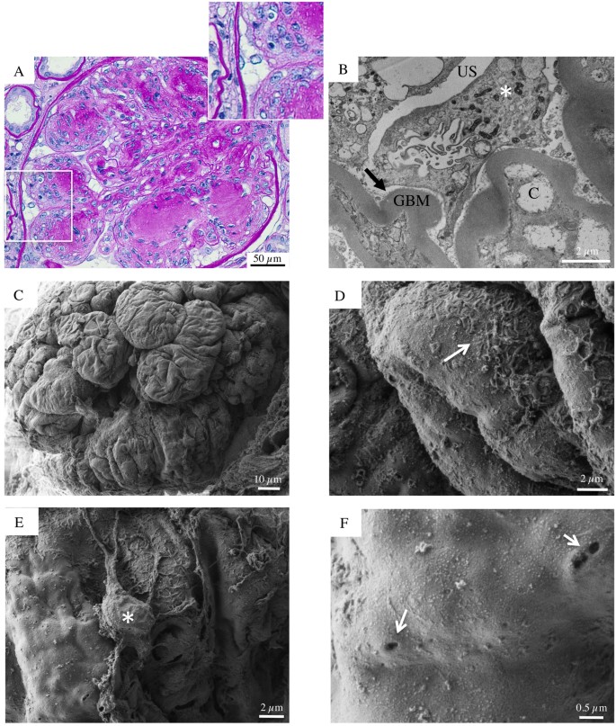
Early and late scanning electron microscopy findings in diabetic kidney  disease | Scientific Reports
Early and late scanning electron microscopy findings in diabetic kidney disease | Scientific Reports
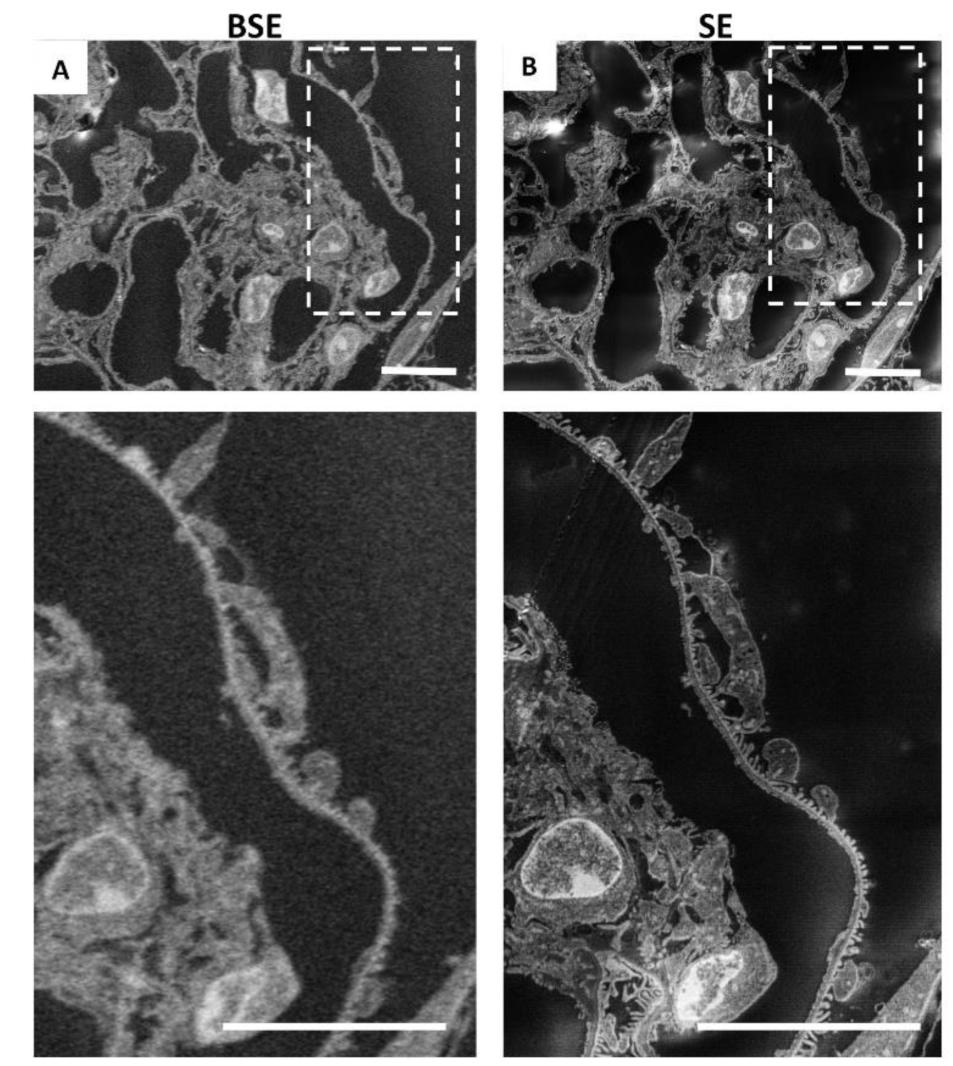
IJMS | Free Full-Text | Imaging the Kidney with an Unconventional Scanning Electron  Microscopy Technique: Analysis of the Subpodocyte Space in Diabetic Mice
IJMS | Free Full-Text | Imaging the Kidney with an Unconventional Scanning Electron Microscopy Technique: Analysis of the Subpodocyte Space in Diabetic Mice

Making Pathology Research More Efficient

JCI Insight - Renal tubular peroxisomes are dispensable for normal kidney  function
JCI Insight - Renal tubular peroxisomes are dispensable for normal kidney function

Here are the key differences between the proximal convoluted tubule (PCT)  and distal convoluted tubule (DCT):- Location: PCT is located immediately  after the glomerulus, while DCT is located more distally in the
Here are the key differences between the proximal convoluted tubule (PCT) and distal convoluted tubule (DCT):- Location: PCT is located immediately after the glomerulus, while DCT is located more distally in the

Transmission electron microscopy image of infected kidney cells of  Piaractus mesopotamicus.
Transmission electron microscopy image of infected kidney cells of Piaractus mesopotamicus.

kidney Dr.Jastrow's electron microscopic atlas

File:Glomerulum of mouse kidney in Scanning Electron Microscope,  magnification 5,000x.GIF - Wikipedia
File:Glomerulum of mouse kidney in Scanning Electron Microscope, magnification 5,000x.GIF - Wikipedia

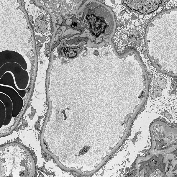
Metabolic Diseases of the Kidney (Chapter 9) - Silva's Diagnostic Renal  Pathology
Metabolic Diseases of the Kidney (Chapter 9) - Silva's Diagnostic Renal Pathology

Figure, Electron Microscopy of Renal Parenchyma...] - StatPearls - NCBI  Bookshelf
Figure, Electron Microscopy of Renal Parenchyma...] - StatPearls - NCBI Bookshelf

Here are the key differences between the proximal convoluted tubule (PCT)  and distal convoluted tubule (DCT):- Location: PCT is located immediately  after the glomerulus, while DCT is located more distally in the
Here are the key differences between the proximal convoluted tubule (PCT) and distal convoluted tubule (DCT):- Location: PCT is located immediately after the glomerulus, while DCT is located more distally in the